Mass Spectrum
Use an MS detector (Mass Spectrometer) to record a mass spectrum at any time t of a chromatogram. This extends the two-dimensional view of a chromatogram (retention time and intensity) by a third dimension (mass). In addition to the information about retention time and intensity, each of the recorded data points includes information about the masses detected in the mass spectrometer.
Mass spectra usually include a great number of narrow peaks or needles. To distinguish them from the comparatively wide chromatogram peaks, they are called mass peaks. A mass spectra representation with many needles is called Centroid. A mass spectra representation that combines the single needles into wide peaks is called a Profile.
In the report, you can display the mass spectrum of a peak or the mass spectrum at any time of the chromatogram. You can also display the current mass spectrum on the control panel during data acquisition.
In addition, you may aggregate single mass spectra to enhance their Signal-to-Noise Ratio (see Using Mass Spectrometers ![]() Processing Mass Spectra).
Processing Mass Spectra).
To display recorded mass spectra on the mass spectra plot, click the following icon on the Method toolbar:
![]()
Mass spectra are generally represented via their relative intensities [%] against the mass. The unit of the mass is m/z (mass/charge number). The base peak, that is, the mass peak with the highest intensity, is the reference point for relative intensities. Thus, its relative intensity is always 100 %.
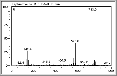
In the above example, the base peak is the mass peak at 733.6 m/z. The time when the mass spectrum was recorded is indicated, also. The mass spectra in the example were aggregated to the displayed total mass spectrum between 0.29 and 0.35 min.
For information about how to record and process MS data, refer to ![]() Using Mass Spectrometers.
Using Mass Spectrometers.